7.0 Tesla MRI Brain Atlas
In Vivo Atlas with Cryomacrotome Correlation
Nonfiction, Health & Well Being, Medical, Specialties, Internal Medicine, Neuroscience, Neurology| Author: | ISBN: | 9781607611547 | |
| Publisher: | Springer New York | Publication: | March 20, 2010 |
| Imprint: | Springer | Language: | English |
| Author: | |
| ISBN: | 9781607611547 |
| Publisher: | Springer New York |
| Publication: | March 20, 2010 |
| Imprint: | Springer |
| Language: | English |
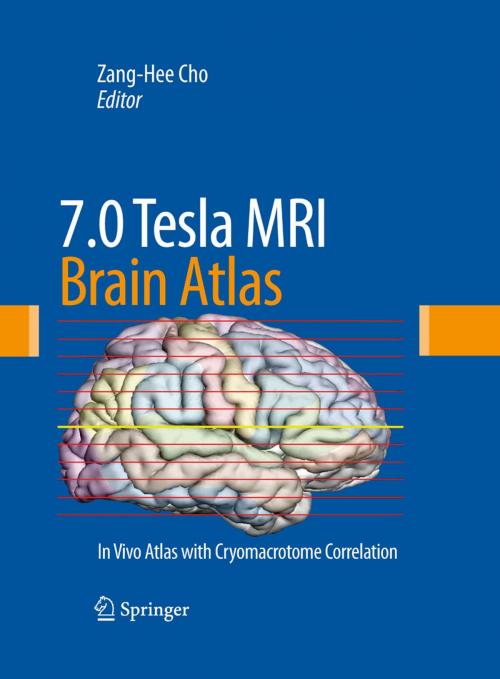
Recent advances in MRI, especially those in the area of ultra high field (UHF) MRI, have attracted significant attention in the field of brain imaging for neuroscience research, as well as for clinical applications. In 7.0 Tesla MRI Brain Atlas: In Vivo Atlas with Cryomacrotome Correlation, Zang-Hee Cho and his colleagues at the Neuroscience Research Institute, Gachon University of Medicine and Science set new standards in neuro-anatomy. This unprecedented atlas presents the future of MR imaging of the brain. Taken at 7.0 Tesla, the images are of a live subject with correlating cryomacrotome photographs. Exquisitely produced in an oversized format to allow careful examination of the brain in real scale, each image is precisely annotated and detailed. The images in the Atlas reveal a wealth of details of the main stem and midbrain structures that were once thought impossible to visualize in-vivo. Ground breaking and thought provoking, 7.0 Tesla MRI Brain Atlas is sure to provide answers and inspiration for further studies, and is a valuable resource for medical libraries, neuroradiologists and neuroscientists.
Recent advances in MRI, especially those in the area of ultra high field (UHF) MRI, have attracted significant attention in the field of brain imaging for neuroscience research, as well as for clinical applications. In 7.0 Tesla MRI Brain Atlas: In Vivo Atlas with Cryomacrotome Correlation, Zang-Hee Cho and his colleagues at the Neuroscience Research Institute, Gachon University of Medicine and Science set new standards in neuro-anatomy. This unprecedented atlas presents the future of MR imaging of the brain. Taken at 7.0 Tesla, the images are of a live subject with correlating cryomacrotome photographs. Exquisitely produced in an oversized format to allow careful examination of the brain in real scale, each image is precisely annotated and detailed. The images in the Atlas reveal a wealth of details of the main stem and midbrain structures that were once thought impossible to visualize in-vivo. Ground breaking and thought provoking, 7.0 Tesla MRI Brain Atlas is sure to provide answers and inspiration for further studies, and is a valuable resource for medical libraries, neuroradiologists and neuroscientists.
