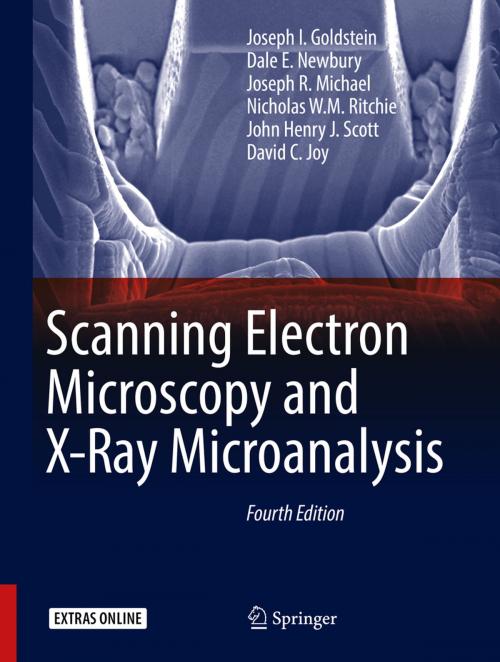
Scanning Electron Microscopy and X-Ray Microanalysis
Nonfiction, Science & Nature, Science, Physics, Spectrum Analysis, Technology, Material Science| Author: | Joseph I. Goldstein, Dale E. Newbury, Joseph R. Michael, Nicholas W.M. Ritchie, John Henry J. Scott, David C. Joy | ISBN: | 9781493966769 |
| Publisher: | Springer New York | Publication: | November 17, 2017 |
| Imprint: | Springer | Language: | English |
| Author: | Joseph I. Goldstein, Dale E. Newbury, Joseph R. Michael, Nicholas W.M. Ritchie, John Henry J. Scott, David C. Joy |
| ISBN: | 9781493966769 |
| Publisher: | Springer New York |
| Publication: | November 17, 2017 |
| Imprint: | Springer |
| Language: | English |
This thoroughly revised and updated Fourth Edition of a time-honored text provides the reader with a comprehensive introduction to the field of scanning electron microscopy (SEM), energy dispersive X-ray spectrometry (EDS) for elemental microanalysis, electron backscatter diffraction analysis (EBSD) for micro-crystallography, and focused ion beams. Students and academic researchers will find the text to be an authoritative and scholarly resource, while SEM operators and a diversity of practitioners — engineers, technicians, physical and biological scientists, clinicians, and technical managers — will find that every chapter has been overhauled to meet the more practical needs of the technologist and working professional. In a break with the past, this Fourth Edition de-emphasizes the design and physical operating basis of the instrumentation, including the electron sources, lenses, detectors, etc. In the modern SEM, many of the low level instrument parameters are now controlled and optimized by the microscope’s software, and user access is restricted. Although the software control system provides efficient and reproducible microscopy and microanalysis, the user must understand the parameter space wherein choices are made to achieve effective and meaningful microscopy, microanalysis, and micro-crystallography. Therefore, special emphasis is placed on beam energy, beam current, electron detector characteristics and controls, and ancillary techniques such as energy dispersive x-ray spectrometry (EDS) and electron backscatter diffraction (EBSD).
With 13 years between the publication of the third and fourth editions, new coverage reflects the many improvements in the instrument and analysis techniques. The SEM has evolved into a powerful and versatile characterization platform in which morphology, elemental composition, and crystal structure can be evaluated simultaneously. Extension of the SEM into a "dual beam" platform incorporating both electron and ion columns allows precision modification of the specimen by focused ion beam milling. New coverage in the Fourth Edition includes the increasing use of field emission guns and SEM instruments with high resolution capabilities, variable pressure SEM operation, theory, and measurement of x-rays with high throughput silicon drift detector (SDD-EDS) x-ray spectrometers. In addition to powerful vendor- supplied software to support data collection and processing, the microscopist can access advanced capabilities available in free, open source software platforms, including the National Institutes of Health (NIH) ImageJ-Fiji for image processing and the National Institute of Standards and Technology (NIST) DTSA II for quantitative EDS x-ray microanalysis and spectral simulation, both of which are extensively used in this work. However, the user has a responsibility to bring intellect, curiosity, and a proper skepticism to information on a computer screen and to the entire measurement process. This book helps you to achieve this goal.
-
Realigns the text with the needs of a diverse audience from researchers and graduate students to SEM operators and technical managers
-
Emphasizes practical, hands-on operation of the microscope, particularly user selection of the critical operating parameters to achieve meaningful results
-
Provides step-by-step overviews of SEM, EDS, and EBSD and checklists of critical issues for SEM imaging, EDS x-ray microanalysis, and EBSD crystallographic measurements
-
Makes extensive use of open source software: NIH ImageJ-FIJI for image processing and NIST DTSA II for quantitative EDS x-ray microanalysis and EDS spectral simulation.
-
Includes case studies to illustrate practical problem solving
-
Covers Helium ion scanning microscopy
-
Organized into relatively self-contained modules – no need to "read it all" to understand a topic
-
Includes an online supplement—an extensive "Database of Electron–Solid Interactions"—which can be accessed on SpringerLink, in Chapter 3


